

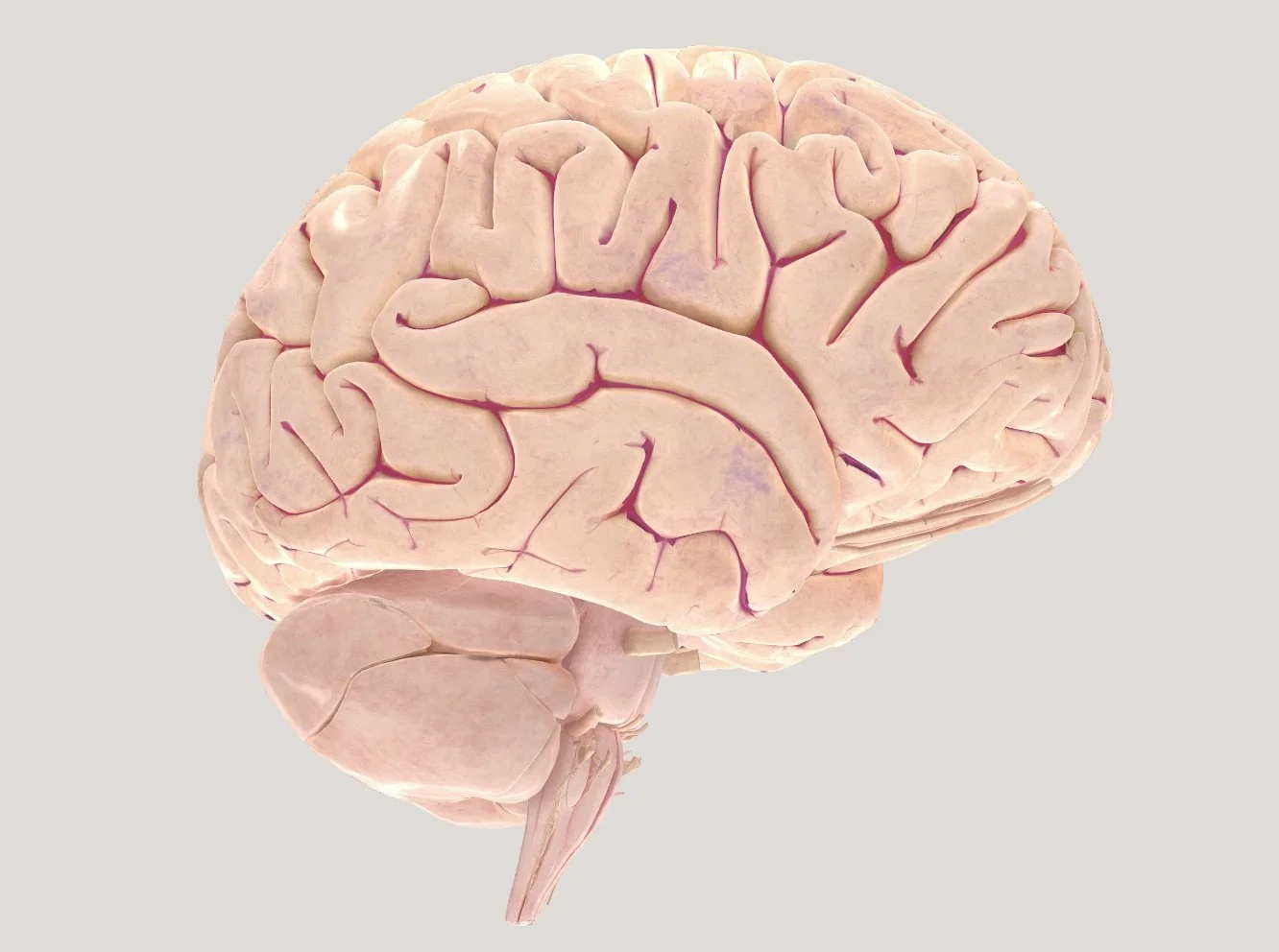
BRAIN ANATOMY AND FUNCTION

THE THREE-POUND UNIVERSE
"It never ceases to amaze me that the brain—a three-pound mass of jelly you can hold in the palm of your hand—can contemplate the vastness of interstellar space, the meaning of infinity, and the mystery of its own existence, all the while trying to figure itself out."
Dr. V.S. Ramachandran in "The Tell-Tale Brain" (2010)
FASCINATING FACTS ABOUT THE HUMAN BRAIN
REALITY IS A BRAIN-CREATED ILLUSION: What we perceive as the external world is actually a complex construct of our brain. Each sensory input—sight, sound, touch—is processed and interpreted in the brain, which then forms an integrated experience we call "reality”.
PEOPLE LIVE PERPETUALLY IN THE PAST: It takes around 80 milliseconds for the brain to process information, so everyone perceives “life” slightly behind real-time.
THE BRAIN HAS 86 BILLION NEURONS, each forming 1000’s of connections with other neurons.
MEMORY CAPACITY: The brain's long-term storage capacity is around 2.5 petabytes, equivalent to about 3 million hours of TV shows.
THE BRAIN CAN REPAIR ITSELF: The brain can reorganise itself by forming new connections, allowing recovery from injury. The brain also changes every time a person learns something new. These processes are called neuroplasticity.
NEGATIVE PLASTICITY: Plasticity is not always positive . The brain can rewire itself in harmful ways, reinforcing bad habits, addictions, or stress responses, especially in those under 25.
PRIMING: Exposure to one stimulus makes a person react faster to something related—like hearing the word "yellow" can make someone think of "bananas."
THE BRAIN HAS NO PAIN RECEPTORS: This allows brain surgeries to be performed on conscious patients.
FASTER THAN A SUPERCOMPUTER: Neurons in the brain can send signals at up to 268 miles per hour, making the brain’s communication network faster than supercomputers.
THE BRAIN IS MORE ACTIVE DURING SLEEP: The brain becomes more active during sleep, consolidating memories and clearing out toxins.
THE BRAIN CONSUMES A LOT OF ENERGY: Although the brain makes up only 2% of a person's body weight, it uses about 20% of the body’s energy and oxygen.
THE BRAIN TAKES 25 YEARS TO FULLY DEVELOP. The frontal lobes responsible for reasoning, decision-making, and impulse control.
THE BRAIN SHRINKS WITH AGE: As a person ages, the brain gradually shrinks. By 60, it may shrink by 0.5-1% per year.
ALIEN HAND SYNDROME: This rare disorder makes one of a person’s hands move on its own, often after brain injury or surgery.
SOME PEOPLE CAN'T RECOGNISE FACES: Prosopagnosia is a condition where a person is unable to recognise faces, even though their vision is normal.
THE BRAIN CAN MAKE PEOPLE THINK THEY'RE DEAD: Cotard's Syndrome makes people believe they are dead or decaying, even though they’re alive and healthy.
SHORT-TERM MEMORY IS EXTREMELY LIMITED: On average, the brain can hold only 5 to 9 items in short-term memory.
PHANTOM LIMB PAIN: Even after amputation, the brain can still feel sensations in missing limbs.
WHERE DID IT ALL BEGIN: THE EVOLUTION OF THE HUMAN BRAIN
4 BILLION YEARS AGO: EARLIEST LIFE FORMS
The earliest forms of life emerged on Earth around 4 billion years ago in the form of protocells—primitive, self-replicating molecules enclosed in membranes. These early life forms eventually evolved into simple multicellular organisms, marking the first steps toward a nervous system.
THE FIRST BRAIN: EARLY VERTEBRATES
Around, 500–600 million years ago, the beginnings of the brain emerged. This can be traced back to early vertebrates, whose nervous systems resembled a simple tube running along the back—like that of the modern lancelet (Amphioxus). This primitive brain wasn’t much more than a swollen segment of the nerve cord, but it already showed signs of specialisation, with nerve cells processing specific information such as light detection and chemical sensing in the water.
As vertebrates evolved, the front part of the nerve cord developed into three distinct sections: the forebrain, midbrain, and hindbrain. This added complexity improved their ability to process increasingly sophisticated information, giving them a survival advantage.

LANCELET (AMPHIOXUS).
THE EVOLUTION OF TETRAPODS AND BRAIN COMPLEXITY
A major turning point in brain evolution occurred when fish evolved into Tetrapods, the first vertebrates capable of crawling out of the water. This shift—from water to land—was driven by selective pressures such as changing habitats and the availability of food and shelter on land. The Tetrapods' evolution gave rise to reptiles, birds, and mammals.
On land, new challenges required more complex sensory processing. The brain regions responsible for sight, sound, and survival behaviours (such as avoiding predators and seeking food) grew significantly. As these functions became more specialised, the forebrain expanded further, eventually forming the right and left hemispheres. Animals that could process complex information from their environment and respond rapidly to threats were more likely to survive and pass on their genes, leading to natural selection favouring those with larger and more intricate brains.
THE RISE OF THE MAMMALIAN BRAIN: CORTEX FOLDING AND HIGHER COGNITION
In early mammals, a remarkable transformation took place: the cortex, the outer layer of the brain, grew in size and complexity. This change didn’t happen randomly—it was a result of mammals needing to process more information as they faced new environmental pressures. Mammals lived in more diverse climates, meaning they had to remember more, solve problems, and engage in social behaviours to survive.
To accommodate the growing number of neurons, the cortex began to fold into ridges and grooves. This folding allowed for a larger surface area, enabling the brain to process more information without needing a larger skull. As mammals became more complex in their behaviours, they developed memory, problem-solving abilities, and social interactions—key features that offered a survival advantage in their environments. The expansion of the cortex and its folding into more intricate patterns enabled the development of advanced cognitive functions like higher-order thinking, language, and abstract reasoning in humans and other mammals.

A TETRAPOD
WHAT IS THE HUMAN BRAIN?
The brain is a highly complex organ that manages every function in our body, from thinking and memory to emotions, movement, vision, breathing, and even regulating temperature and hunger; everything from essential bodily functions to advanced cognitive processes like thinking and problem-solving. The brain works closely with the spinal cord, forming the central nervous system (CNS), which controls the body’s vital processes.
WHAT IS THE BRAIN MADE OF?
The average adult brain weighs about three pounds and comprises roughly 60% fat, with the remaining 40% being water, protein, carbohydrates, and salts. Despite its complexity, the brain is not a muscle. It contains blood vessels and nerves, including specialised nerve cells called neurons and supportive cells known as glial cells.
WHAT IS GRAY MATTER AND WHITE MATTER?

The brain and spinal cord contain two types of tissue: gray matter and white matter.
Gray matter refers to the darker outer layer of the brain, consisting mainly of neuron somas (the central cell bodies of neurons).
White matter is the lighter inner region beneath the gray matter. It consists of axons (long neuron tails) covered in myelin, a fatty protective layer (mylination does not finish until the mid twenties).
In the spinal cord, the arrangement is reversed: white matter is on the outside, and gray matter is found on the inside. The different structures of neurons in each matter give them their distinctive colours in medical scans.
Gray matter is responsible for processing and interpreting information.
White matter acts as the brain’s communication network, transmitting information between different parts of the nervous system.
HOW DOES THE BRAIN WORK?

The brain communicates by sending and receiving chemical and electrical signals throughout the body. These signals control everything from mood and energy levels to pain and other sensations. Some signals stay within the brain, while others travel through the spinal cord and are transmitted via nerves to different body parts.
The central nervous system relies on billions of specialised cells called neurons to relay these messages, allowing the brain to coordinate everything we do, think, and feel.
INTRODUCTION TO BRAIN ANATOMY
This section will explore the brain's organisation, including its significant regions and structures, and how these different parts contribute to its overall function. We'll look at how the brain is divided into key sections, such as the forebrain, midbrain, and hindbrain, and discuss the significance of the cerebral cortex, which plays a vital role in higher-level thinking. Understanding the brain's structure is crucial for grasping how it controls voluntary and involuntary bodily processes.
BRAIN PARTS AND THEIR FUNCTION
The brain is divided into three main parts: the hindbrain, midbrain, and forebrain. These regions developed at different stages of evolution, each contributing to essential functions necessary for survival and higher cognitive processes.

HINDBRAIN:
This is the most primitive part of the brain, found in early vertebrates. It consists of four parts: the medulla, reticular formation, cerebellum, and pons.
It primarily regulates necessary functions such as breathing, heart rate, and motor control. It is located at the base of the brain, near the top of the spinal cord.
BRAINSTEM
The brainstem, located in the middle of the brain, it connects the cerebrum to the spinal cord.
PONS
The pons is located above the medulla and below the midbrain, as part of the brainstem.
It bridges different parts of the nervous system, including the cerebellum and cerebrum. The pons also regulate sleep, breathing, and facial expressions.
MEDULLA
The medulla is found at the base of the brainstem.
It controls vital automatic functions such as breathing, heart rate, and blood pressure. Damage to the medulla can be life-threatening as it regulates functions essential for survival.
RETICULAR FORMATION
The reticular formation is a network of nerves that runs through the brainstem, including the medulla and pons.
It is crucial for attention and focus, helping to filter and prioritise sensory stimuli. It allows a person to focus on essential stimuli while filtering out less meaningful distractions. For example, in a dangerous situation, the reticular formation helps the person focus solely on survival.
During sleep, the reticular formation slows down. Still, it continues to filter sensory input, so a person might sleep through ordinary sounds like traffic but wake up to an alarm or smoke detector.
CEREBELLUM: THE "LITTLE BRAIN"
The Cerebellum is Situated at the base of the brain, beneath the occipital lobe and behind the brainstem in a space known as the posterior cranial fossa.
The Cerebellum is known as the "little brain," because it looks like a mini version of the Cerebrum, for example, s it has two hemispheres and a highly folded surface (cortex). However, its functions and evolutionary history differ significantly from the cerebrum.
The cerebellum coordinates unconscious movements, such as balance, posture, and fine motor control. It ensures that movements are smooth and precise. While it does not initiate movement, it plays a critical role in the timing and accuracy of movements initiated by other brain parts, particularly the motor cortex.
The cerebellum helps store learned motor patterns. Tasks such as playing an instrument or riding a bike, which initially require conscious effort, become automatic with practice. Once these movements are learned, the cerebellum coordinates them without conscious thought, refining and smoothing out complex motor skills. For example, in London, taxi drivers who have mastered "The Knowledge" and their ability to navigate routes automatically rely on motor patterns stored in the cerebellum.
CONNECTION TO THE AUTONOMIC NERVOUS SYSTEM: The cerebellum is not directly connected to the autonomic nervous system (ANS), which regulates involuntary functions like heart rate and digestion. However, its role in maintaining balance and posture can indirectly influence specific autonomic processes, such as stabilising breathing during movement or physical activity.
EFFECTS OF DAMAGE TO THE CEREBELLUM: Damage to the cerebellum can significantly impair movement and coordination. The following issues may arise:
Ataxia: A loss of coordination that affects balance and gait, making walking or standing difficult.
Tremors: Shaky, unsteady movements, especially noticeable when performing intentional tasks like reaching for an object.
Difficulty with Balance and Posture: Problems maintaining balance or proper posture, leading to instability when sitting or walking.
Impaired Fine Motor Control: Tasks requiring precise, controlled movements, such as writing, typing, or buttoning clothes, become challenging and clumsy.
EVOLUTIONARY CONTEXT
The cerebellum is considered evolutionarily older than the cerebrum. This means it likely developed earlier in the evolutionary timeline. Many animals, such as reptiles, have a well-developed cerebellum but lack a cerebral cortex, which is a more recent evolutionary development seen in mammals and especially primates. This suggests that the cerebellum plays a fundamental role in movement and survival functions, which were essential for early vertebrates long before higher cognitive functions (associated with the cerebral cortex) evolved.
Evolution: The hindbrain is the most ancient part of the brain, found in all vertebrates, including reptiles, birds, and mammals. It is essential for basic life processes like survival and reflexes. This part evolved over 500 million years ago, appearing first in early fish.

THE MIDBRAIN:
Location: Situated above the hindbrain, forming the central part of the brain.
Function: It is an essential connection between the brain's lower and higher areas, relaying sensory and motor information. It also plays a role in vision, hearing, and motor control, contributing to arousal and alertness.
Relay Station: The midbrain acts as a relay station for sensory and motor information, transmitting signals between the forebrain and spinal cord. It ensures that sensory data, such as visual and auditory signals, reach the appropriate parts of the brain.
Vision and Hearing: The midbrain plays a crucial role in processing visual and auditory information. It helps control eye movement and pupil dilation in response to light and processes sounds to determine their source and significance.
Movement Control: The midbrain helps regulate motor movement, particularly involuntary movements. It contains structures like the substantia nigra, which are involved in the control of voluntary movement and are linked to disorders such as Parkinson’s disease when damaged.
EXAMPLES
When you hear a loud sound, your midbrain helps orient your head and eyes toward the source.
The midbrain also helps coordinate movements such as walking and maintaining posture.
Evolution: The midbrain evolved after the hindbrain approximately 400 million years ago. In reptiles and amphibians, the midbrain handles much of their sensory processing. However, the midbrain’s role is reduced in mammals as the forebrain takes over more complex functions.
THE FOREBRAIN:
Location: The largest and most advanced part of the brain, found at the top and front.
Function: FOREBRAIN: The largest and most advanced part of the brain, the forebrain, developed last in evolutionary terms. It encompasses the cerebrum, which includes the cerebral cortex and subcortical structures like the thalamus and hypothalamus. The forebrain is responsible for higher cognitive functions such as thought, decision-making, emotions, and voluntary movement.
Evolution: The forebrain is the most recent part of evolution, developing around 200 million years ago in early mammals. This part supports advanced cognition, especially in humans, primates, and dolphins.
THE PINEAL GLAND
Pineal Gland—located deep in the center of the brain, in the midline between the two hemispheres; part of the endocrine system; produces melatonin, which helps maintain circadian rhythm and regulate reproductive hormones; also regulates other endocrine functions and converts signals from the nervous system into endocrine signals
THE THALAMUS
The thalamus acts as a relay station, processing sensory signals (such as touch, sight, and sound) and sending them to the relevant parts of the cerebral cortex for interpretation.
Example: When you hear a sound, the thalamus passes this information to the cortex to help you recognise it, like identifying a friend’s voice.
THE HYPOTHALAMUS:
The hypothalamus regulates homeostasis, maintaining the body’s internal balance (e.g., temperature, hunger, thirst, and sleep). It also influences emotional responses and the endocrine system, which controls hormone production.
LOCATION: The hypothalamus is located deep within the brain, below the thalamus and above the pituitary gland. It is a small but vital structure, forming part of the diencephalon and situated near the base of the brain.
The hypothalamus connects directly to the pituitary gland via a stalk known as the infundibulum, which is crucial in linking the nervous system to the endocrine system.
FUNCTION: The hypothalamus regulates many of the body’s essential processes. It acts as a control centre for the autonomic nervous system (ANS) and endocrine system, making it critical for maintaining homeostasis (the body's internal balance).
Some of its other functions include:
Regulation of body temperature: The hypothalamus monitors and adjusts body temperature, prompting actions like sweating or shivering when necessary.
Control of hunger and thirst: It signals when the body needs food or water and regulates eating and drinking behaviour.
Sleep-wake cycle (circadian rhythms): The hypothalamus helps regulate sleep patterns by controlling the release of melatonin and coordinating with the pineal gland.
Hormone release: Through interaction with the pituitary gland, the hypothalamus governs the release of hormones that regulate growth, metabolism, and reproduction. This includes corticotropin-releasing hormone (CRH), which activates the HPA axis during stress.
Emotion and behaviour: The hypothalamus is involved in emotional responses, including feelings such as fear, anger, and pleasure. It also helps control behaviours like aggression and sexual activity.
EFFECTS OF HYPOTHALAMIC DAMAGE
Damage to the hypothalamus can have wide-ranging effects due to its control over many vital functions. Some of the potential consequences include:
Body temperature dysregulation: Damage may cause the body to have difficulty maintaining a stable temperature, leading to hypothermia or hyperthermia.
Appetite and thirst disorders: Damage to the hypothalamus can result in excessive hunger or thirst (or the opposite, leading to a loss of appetite) and conditions like diabetes insipidus (a condition that affects water balance).
Sleep disturbances: Injuries to the hypothalamus can disrupt standard sleep patterns, causing insomnia or hypersomnia (excessive sleep).
Hormonal imbalances: Since the hypothalamus governs the release of many hormones through the pituitary gland, damage can lead to hormonal problems, affecting growth, reproduction, stress response, and metabolism.
Emotional and behavioural changes: Damage to the hypothalamus can also result in unusual emotional responses, such as extreme aggression or apathy, due to its role in regulating emotional behaviour.
Difficulty regulating stress: Since the hypothalamus activates the HPA axis during stress, damage may impair the body's ability to manage stress effectively
THE CEREBRUM:
The cerebrum is contained with the forebrain. It is the most significant part of the brain and handles higher cognitive functions like thinking, decision-making, and voluntary movements.
LATERALISATION
The cerebral cortex is split into two halves, or hemispheres, which are covered with ridges (gyri) and grooves (sulci). These two halves are separated by a deep groove called the inter hemispheric fissure, also known as the medial longitudinal fissure, which runs from the front to the back of the brain. The right hemisphere controls the left side of the body, while the left hemisphere controls the right side. Both hemispheres communicate via a large, C-shaped bundle of white matter and nerve fibres called the corpus callosum, located in the centre of the cerebrum.
CORPUS CALLOSUM:
This large bundle of nerve fibres connects the left and right hemispheres of the brain, allowing them to communicate with each other. It plays a key role in coordinating activities between both sides of the brain.
LOBE AND CORTICES

.
The cerebrum is divided into divided four brain lobes. The word "lobe" comes from the Greek "lobos," meaning "a small rounded projection that appears separate from the rest due to a fissure (crack), sulcus (groove), connective tissue, or shape.
THE FOUR LOBES ARE:
Frontal Lobe
Parietal Lobe
Temporal Lobe
Occipital Lobe
These lobes manage broad functions like sensory perception, movement, and complex thinking. Within each lobe are smaller specialised areas called cortices that handle specific tasks like processing vision, generating speech, or controlling movement.
Let’s explore these regions in more detail.
THE FRONTAL LOBE
Location: The frontal lobe is at the front of the cerebrum, just behind the forehead.
The frontal lobe is responsible for cognitive thinking and significantly influences an individual's personality. It typically reaches maturity by age 25, when cognitive maturity is attained. Functions of the frontal lobe include reasoning, planning, behaviour regulation, emotions, problem-solving, and motor skills. It also plays a role in speech production.
EFFECTS OF FRONTAL LOBE DAMAGE
Due to its location at the front of the skull, the frontal lobe is particularly vulnerable to injury, which may result in:
Altered talking habits
Impaired judgement and risk-taking ability
Loss of taste or smell
Increased distractibility
Memory loss and dementia, which can contribute to conditions like Alzheimer's disease
Critical Areas in the Frontal Lobe:
Broca’s Area: Responsible for language production. Damage to this area can result in Broca’s Aphasia, an inability to produce language.
Prefrontal Cortex: Essential for higher-order cognitive functions, such as problem-solving, emotional regulation, and complex thought.
Associated Cortex: The prefrontal cortex is located in the front of the frontal lobe and responsible for decision-making, impulse control, and planning. The primary motor cortex is located at the rear of the frontal lobe, along the precentral gyrus, and controls voluntary muscle movements.
THE TEMPORAL LOBE
Location: Located on the sides of the cerebrum, roughly behind the ears.
The temporal lobe contains the primary auditory cortex, which is responsible for auditory processing. It also houses the hippocampus, which is crucial in forming long-term memories. The right temporal lobe is responsible for visual memory, while the left manages verbal memory.
THE FUSIFORM FACE AREA (FFA)
LOCATION: The fusiform face area (FFA) is located in the fusiform gyrus, a part of the brain's temporal lobe. Specifically, it is found on the brain's ventral (bottom) surface within the occipitotemporal cortex. The FFA is primarily located in the right hemisphere, although a corresponding region in the left hemisphere may also be involved to some extent.
FUNCTION: The FFA is specialised for recognising and processing faces. It helps us identify familiar faces and distinguish between different individuals. This area plays a crucial role in our ability to:
Recognise facial features
Detect facial expressions
Understand emotional cues from faces
Differentiate between faces and other objects
While the FFA is particularly sensitive to faces, it may also contribute to expertise in other areas, such as recognising objects in fields where individuals have developed high expertise (e.g., birdwatchers identifying birds, car experts recognising car models).
WHAT HAPPENS IF THE FFA IS DAMAGED?
Damage to the fusiform face area can lead to prosopagnosia, also known as face blindness. People with prosopagnosia have difficulty recognising faces, even those of family and close friends. However, they may still be able to recognise individuals by other cues like voice, clothing, or hairstyle.
In summary, the fusiform face area plays a critical role in facial recognition and contributes to interpreting social and emotional information from faces.
EFFECTS OF TEMPORAL LOBE DAMAGE
Left Temporal Lobe Damage: Difficulty recognising words, remembering verbal material, and recalling audio and visual content.
Right Temporal Lobe Damage: Difficulty recognising faces (prosopagnosia), tonal sequences, and recalling music or drawings.
General Effects: Damage can lead to short-term memory loss, aggressive behaviour, difficulty with speech comprehension (Wernicke’s Aphasia), and increased talkativeness.
Associated Cortices: The auditory cortex is located in the upper part of the temporal lobe, near the ears, and processes sound. Wernicke’s area is located in the left temporal lobe and is crucial for understanding language.
THE PARIETAL LOBE
Location: The parietal lobe sits near the top and back of the cerebrum, behind the frontal lobe.
The parietal lobe integrates sensory information from the body, such as touch, pain, and temperature. It also plays a role in spatial orientation and visual perception.
Critical Functions of the Parietal Lobe:
Recognising spatial relationships, such as determining where tactile sensations are coming from.
They discriminate the intensity of stimuli, such as distinguishing between cold and ice-cold.
EFFECTS OF PARIETAL LOBE DAMAGE
Left Parietal Lobe Damage may result in Gerstmann’s syndrome, aphasia, and agnosia (inability to recognise objects).
Right Parietal Lobe Damage: Impairs personal care skills, drawing ability, and spatial processing.
Bilateral Parietal Lobe Damage: Causes Balint’s syndrome, which impairs visual attention and motor control.
Associated Cortex: The somatosensory cortex is located in the front part of the parietal lobe, along the postcentral gyrus (just behind the central sulcus), and processes sensations like touch and pain from the body
THE OCCIPITAL LOBE
Location: Found at the very back of the cerebrum.
The occipital lobe is the smallest of the brain’s lobes and is responsible for visual perception. The primary visual cortex processes visual information such as colour, motion, and spatial relationships.
EFFECTS OF OCCIPITAL LOBE DAMAGE
Visual Field Cuts: Loss of vision in specific areas.
Colour Agnosia: Difficulty recognising colours.
Movement Agnosia: Inability to perceive movement.
Word Blindness: Inability to recognise written words.
Associated Cortex: The primary visual cortex is located in the rear part of the occipital lobe and processes raw visual data received from the eyes..
THE CEREBRAL CORTEX
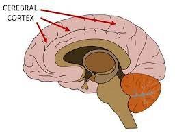
The cerebral cortex is essential for everything that makes us human—from language and reasoning to creativity and emotional intelligence. Without it, we couldn't engage in complex thought, make plans, or experience a rich inner life; making it the "crowning glory" of the cerebrum.
LOBE VS CORTEX: WHAT’S THE DIFFERENCE?
The cerebral cortex is the cerebrum's outermost layer, the brain's largest part. The word "cortex" comes from the Latin word for "bark," as it forms a thin (about 1/4-inch thick) layer that covers the entire surface of the cerebrum.
This highly folded structure allows for more surface area, meaning more neurons, which increases the brain’s capacity for advanced functions. Ultimately, the cerebral cortex covers all of the lobes.
But just as the cerebrum is divided into four lobes the cerebral cortex is also divided into sections. These sections correspond to specific areas of the lobes and have distinct functions known as cortices. For instance, the motor cortex, located within the frontal lobe, controls voluntary movement.
This distinction highlights how the brain is organised: the lobes manage broader functional categories, while the cortices within them focus on more specific, task-oriented roles. You can think of the lobes as large sections of the brain responsible for general functions, with the cortices as specialised areas within these sections, performing precise tasks

HOW THEY WORK TOGETHER
While lobes are broader regions, the cortices within them work together to ensure that more complex tasks are carried out efficiently. For instance, the frontal lobe is associated with movement, reasoning, and personality. Still, specific cortices within the frontal lobe handle particular aspects of these functions, such as motor control or language production.
TYPES OF CORTEX
PREFRONTAL CORTEX (FRONTAL LOBE)

LOCATION: The prefrontal cortex is located at the front of the frontal lobe, just behind the forehead.
FUNCTION: This brain region is crucial for higher cognitive functions such as planning, decision-making, problem-solving, and impulse control. It allows us to anticipate future consequences, manage complex social interactions, and control our emotions. The prefrontal cortex also involves personality expression and behaviour regulation, ensuring that actions align with goals and social norms. Damage to this area can lead to poor judgement, impulsivity, and difficulty in managing emotions, as it plays a central role in governing how we make thoughtful decisions and manage our impulses.
FRONTAL CORTEX (FRONTAL LOBE)

LOCATION: The frontal cortex encompasses the entire frontal lobe, including the prefrontal cortex, and extends from behind the forehead to the central sulcus, where it meets the parietal lobe.
FUNCTION: The frontal cortex is responsible for various functions, including voluntary movement, cognitive skills, and language processing. It governs our ability to think, plan, and execute complex behaviours. While the prefrontal cortex handles executive functions, the motor areas of the frontal cortex control the execution of movements. This region is also involved in speech production via Broca's area. The frontal cortex shapes our behaviour, coordinating motor control and higher cognitive functions essential for everyday life.
MOTOR CORTEX (FRONTAL LOBE)

LOCATION: Located in the frontal lobe, along the precentral gyrus, just in front of the central sulcus.
FUNCTION:
The primary motor cortex controls voluntary movements. Different regions correspond to other body parts, such as the hands, feet, and face, helping coordinate muscle movement.
SOMATOSENSORY CORTEX (PARIETAL LOBE)

SOMATOSENSORY CORTEX
LOCATION:
It is situated in the parietal lobe, behind the primary motor cortex on the postcentral gyrus.
FUNCTION:
The somatosensory cortex processes sensory information from the body, including touch, pressure, pain, and temperature. It helps the brain understand where limbs are in space and how they interact with objects.
VISUAL CORTEX (OCCIPITAL LOBE)

LOCATION:
It is found in the occipital lobe, at the back of the brain.
FUNCTION:
The visual cortex processes visual information from the eyes, allowing you to recognise shapes, colours, motion, and depth.
AUDITORY CORTEX
The auditory cortex is located in the temporal lobe, near the ears on both sides of the brain.
FUNCTION: THE AUDITORY CORTEX: Detection of Sound Quality - Loudness, Tone, Volume, Tempo, Pitch.
The auditory cortex is crucial for analysing and processing acoustic information, dealing with various sound dynamics such as volume, tempo, and pitch. It handles the initial sounds we hear and, through a process called parallel distributed processing, sends different aspects of the sounds to various parts of the brain for further processing—much like an assembly line in a factory. The auditory cortex is bilaterally organised, meaning it exists in both temporal lobesof the brain. This bilateral structure enhances spatial awareness and allows for a more comprehensive interpretation of sound.
While the auditory cortex processes basic sound features like loudness, tone, volume, tempo, and pitch, the content of the sound—what the sound actually is—is processed differently. This type of information is sent to specific areas of the brain for specialised processing, creating some lateralised specialities within the auditory system.
For example, verbal information—such as native languages, second languages, nonsense words, and even backwards speech—is processed predominantly in the left hemisphere. The area responsible for this is known as Wernicke’s area, located primarily in the left temporal lobe. Its function is to handle the semantics of speech, meaning it helps us understand the meaning behind the sounds. If Wernicke’s area is damaged, a person can still hear speech but will struggle to comprehend what is being said, demonstrating that its role is not in interpreting sound but in decoding meaning.
In contrast, non-verbal sounds—such as animal noises, music, and onomatopoeia—tend to be processed with a right-brain advantage. The right hemisphere specialises in interpreting emotional tones in speech (intonation) and in recognising environmental and musical sounds.
This lateralisation likely evolved to aid survival, enabling early humans to distinguish important sounds, such as animal calls or environmental noises, from human speech. This would have helped them differentiate between predator and prey sounds during hunting
LOCATION: Situated in the temporal lobe, close to the ears.
FUNCTION: The auditory cortex processes raw sound information, including pitch, volume, and rhythm, and helps to identify different types of sound, like speech, music, and environmental noises.
WERNICKE’S AREA (LANGUAGE COMPREHENSION)

WERNICKE’S AREA
.
LOCATION: Located in the left temporal lobe, near the auditory cortex.
FUNCTION: Wernicke’s area is responsible for understanding language and comprehending spoken words. After the auditory cortex processes the sound, Wernicke’s area interprets the meaning of the sounds, especially in terms of language (e.g., recognising words and making sense of sentences). Damage to this area results in Wernicke's aphasia, where a person can speak fluently but has difficulty understanding spoken language, and their speech may lack meaningful content.
BROCA’S AREA (LANGUAGE PRODUCTION)

BROCAS AREA
.
LOCATION: Found in the left frontal lobe, near the motor cortex (closer to the front of the brain).
FUNCTION: While Broca’s area is not in the temporal lobe, it works closely with the auditory cortex and Wernicke’s area for speech production. After Wernicke’s area interprets the meaning of words, Broca’s area helps to form the motor plans necessary to produce speech. Damage to Broca’s area results in Broca's aphasia, where an individual understands language but struggles to produce speech fluently.
HOW THEY WORK TOGETHER
Auditory Cortex: Processes the sound of spoken words, music, and other noises, e.g., their pitch, volume, etc..
Wernicke’s Area: Interprets the meaning of these words (language comprehension).
Broca’s Area: Creates the motor instructions to produce speech (language production).
In short, the auditory cortex handles the primary processing of sound, while Wernicke’s and Broca’s areas handle understanding and producing language. They aren’t subdivisions of the auditory cortex but work together for speech and language-related tasks.
WHY IS THE CEREBRAL CORTEX WRINKLY?
One of the cerebral cortex's most notable features is its wrinkled surface, caused by cortical folding. The brain's surface comprises gyri (ridges) and sulci (grooves), which increase its surface area. This folding allows the brain to fit more neurons (brain cells) within the limited space of the skull, enhancing its ability to process information efficiently. The more wrinkled the cortex, the more neurons it can hold, supporting advanced functions like reasoning, memory, and learning.
To illustrate this, imagine fitting a large bed sheet into a small purse. You would need to fold it several times to make it fit. Similarly, the brain folds its cortex to maximise space, enabling more excellent cognitive capabilities. Over two-thirds of the cortex is buried within the sulci, showing how vital this folding is for brain efficiency.
SULCI, GYRI, AND BRAIN ANATOMY
The names of these folds and wrinkles are:
Gyri (singular: gyrus) are the raised ridges on the brain's surface.
Sulci (singular: sulcus) are the grooves or indentations between the gyri.
This pattern of gyri and sulci is unique to each individual but follows a general layout across humans, helping to identify specific brain regions and their functions. For example, the precentral gyrus is involved in motor control, while the postcentral gyrus is essential for processing somatosensory information like touch, pressure, and temperature.

IMPORTANT GYRI AND SULCI
Each gyrus and sulcus corresponds to specific brain functions. They help us identify where different functions happen in the brain. Here are a few examples:
Precentral Gyrus is in the frontal lobe, just before the central sulcus. It’s the home of the primary motor cortex, which controls voluntary movement like picking up a cup or kicking a ball.
Postcentral Gyrus: Found just behind the central sulcus in the parietal lobe, it contains the somatosensory cortex, where the brain processes touch, temperature, and pain.
Lateral Sulcus (or Sylvian Fissure): This groove separates the temporal lobe (responsible for hearing and language) from the frontal and parietal lobes.
Parieto-occipital Sulcus separates the parietal lobe from the occipital lobe, where visual processing happens.
FISSURES
Fissures are deeper grooves that divide the brain into different sections, e.g., a narrow slit or aperture, especially one of the deeper or more constant furrows separating the gyri of the brain.
The longitudinal fissure runs down the middle of your brain, dividing it into left and right hemispheres.
The transverse fissure separates the cerebrum (the largest part of the brain) from the cerebellum (the part that helps with movement and balance).
HOW GYRI AND SULCI HELP
The pattern of gyri and sulci is like a brain map, showing us where different functions are located.
EVOLUTIONARY COMPARISONS: ANIMAL BRAIN STRUCTURES
This intricate folding is a hallmark of mammalian brains, This folding is much more pronounced in humans and other intelligent animals. For example, primates and dolphins also have highly folded brains, while simpler animals like reptiles have smooth brains with fewer cognitive abilities.
Animals that lack a cerebral cortex, like reptiles, don’t have complex cognitive abilities like humans. They don’t recognise emotions, understand social interactions, or have self-awareness.
REPTILES:
Brain Structure: Reptiles, like lizards and snakes, have relatively small forebrains and lack a developed limbic system (which handles emotions). As a result, they show little to no emotional responses and rely heavily on instinct for survival.
Cognitive Abilities: Reptiles lack theory of mind (the ability to understand others’ thoughts and emotions) and self-awareness. They don't recognise faces or process emotions like humans or other mammals. Their behaviour is driven by survival instincts, such as hunting or finding shelter, without social bonding or emotional depth.
BIRDS:
Birds have a more developed forebrain than reptiles, but they lack a cerebral cortex, which is critical for higher cognitive functions in mammals. Instead, birds rely on a structure called the pallium for problem-solving.
Cognitive Abilities: Birds like crows and parrots are brilliant and capable of tool use and mimicry, but they do not exhibit the emotional recognition or self-awareness seen in mammals. They can solve complex problems but don’t have speech or face recognition abilities like humans.

Make it stand out
SUMMARY: LOBES AND CORTICES
Lobes are the broad regions of the cerebrum that manage general functions like movement, sensation, and thought.
Cortices are specialised areas within the lobes that handle specific tasks, such as processing sensory input, controlling speech, or enabling voluntary movement.
The cerebral cortex is the brain’s outer layer and covers all lobes. It is highly wrinkled to increase surface area for complex thinking and processing.
The wrinkly cerebral cortex is key to intelligence. Humans, primates, and dolphins have the most wrinkled cortices, which enable complex thoughts and actions.
DEEPER STRUCTURES WITHIN THE BRAIN

THE MESOLIMBIC SYSTEM: AN OVERVIEW
The Mesolimbic system, often referred to as the reward pathway, is a complex network of brain structures that plays a vital role in motivation, pleasure, and behaviour reinforcement. This system is central to the brain’s ability to experience pleasure and is activated by rewarding stimuli such as food, social interactions, and addictive substances. When the reward pathway is stimulated, it reinforces the likelihood that a behaviour will be repeated by activating key regions of the brain. Here is a breakdown of the key components:
KEY COMPONENTS OF THE MESOLIMBIC SYSTEM
VENTRAL TEGMENTAL AREA (VTA):
Location: The VTA is located in the midbrain.
Function: The VTA is the origin of dopamine neurons that project to various parts of the brain, including the nucleus accumbens. It is responsible for the initial release of dopamine, which is the key neurotransmitter in the reward pathway. This release of dopamine signals pleasure and reward, motivating behaviour.
Role in Addiction: Addictive drugs stimulate dopamine release from the VTA, making this structure critical in addiction and compulsive behaviour.
NUCLEUS ACCUMBENS (NA):
Location: The nucleus accumbens is located in the basal forebrain, near the head of the caudate nucleus.
Function: The nucleus accumbens is the primary site where dopamine from the VTA is released. This area is crucial for the experience of pleasure and reinforcement of rewarding behaviour. It is sometimes referred to as the "pleasure centre" of the brain.
Role in Motivation: When dopamine is released in the nucleus accumbens, it creates a sense of pleasure or reward, which motivates individuals to repeat the action that triggered the release.
AMYGDALA:
Location: The amygdala is deep within the temporal lobe and part of the limbic system.
Function: The amygdala processes emotions, including fear and pleasure. It helps the brain associate emotions with experiences, critical in learning and memory. The amygdala contributes to emotional reinforcement in the reward system by allowing the brain to remember which behaviours lead to pleasurable experiences.
Role in Reward and Fear: While the amygdala is heavily involved in fear responses, it also plays a role in emotional learning related to rewards, helping to solidify behaviours that result in pleasure.
HIPPOCAMPUS:
Location: The hippocampus is beneath the cortical surface in the medial temporal lobe.
Function: The hippocampus is essential for forming long-term memories. In the context of the mesolimbic system, it helps store memories of past rewards and reinforces behaviour based on the memory of pleasure. This ensures that actions leading to positive experiences are remembered and repeated.
Role in Addiction: The hippocampus plays a role in remembering the context of rewarding experiences, such as where and when pleasurable activities occurred, making it relevant in habits and addictions.
PREFRONTAL CORTEX (PFC):
Location: The PFC is located in the frontal lobes at the front of the brain.The main functions of the have already been discussed above.
But in the mesolimbic system, the PFC helps regulate responses to rewards and assess long-term consequences of actions. Given future implications, it allows individuals to weigh whether an immediate reward is worth pursuing.
Role in Addiction: Dysfunction in the PFC is often linked to addictive behaviour, as it impairs the ability to resist short-term rewards despite long-term adverse outcomes.
STRIATUM:
Location: The striatum is part of the basal ganglia and deep within the cerebral hemispheres.
Function: The striatum coordinates motor activity and reward processing. It is involved in translating motivation into action. Dopamine release in the striatum motivates physical actions to seek out rewards.
Role in Habit Formation: The striatum is critical in forming habits and reinforcing behaviours that have previously led to rewards, making it a crucial part of compulsive behaviour and addiction.
HOW THE MESOLIMBIC SYSTEM WORKS
The mesolimbic system operates as a circuit that starts with the ventral tegmental area (VTA), which releases dopamine. The dopamine travels to key structures like the nucleus accumbens and the prefrontal cortex, creating feelings of pleasure, motivation, and reinforcement. As these areas communicate with the amygdala and hippocampus, memories of the rewarding experience are stored, making the behaviour more likely to be repeated.
OTHER IMPORTANT STRUCTURES DEEP WITHIN THE BRAIN
PITUITARY GLAND: Often called the "master gland," the pituitary gland is a small, pea-sized structure located deep in the brain behind the bridge of the nose. It controls other glands in the body, such as the thyroid, adrenals, ovaries, and testicles, by regulating hormone release. The pituitary receives chemical signals from the hypothalamus, which guides its functions.
CAUDATE NUCLEUS: Part of the basal ganglia, the caudate nucleus is involved in motor control, learning, and habit formation. It also processes information related to rewards and motivation.
These deeper structures regulate essential body functions, from hormonal balance and sleep to movement, memory, and emotions.
ANATOMICAL TERMS OF LOCATION IN BRAIN STRUCTURE

.
CRANIAL, CAUDAL, AND ROSTRAL
Specific terms exist to describe how close or far something is to the head or tail of an animal. To explain how close to the head of an animal something is, three distinct terms are used:
Rostral, from Latin rōstrum ("beak, nose"): situated toward the oral or nasal region, or in the case of the brain, toward the tip of the frontal lobe.
Cranial, from Greek κρᾱνίον (kranion, "skull") or cephalic (κεφαλή (kephalē, "head").
SUPERIOR AND INFERIOR
In anatomical terminology, superior refers to a structure above another, while inferior means below another structure.
An example in the brain is that the cerebral cortex is superior to the brainstem because it is located above it. Similarly, the cerebellum is inferior to the occipital lobe of the cerebrum because it is located below it.
ANTERIOR AND POSTERIOR
Anterior refers to structures toward the front of the body or brain, and posterior refers to structures toward the back.
Example in the brain: The frontal lobe of the cerebrum is anterior to the parietal lobe, as it is positioned at the front of the brain. Conversely, the occipital lobe is posterior to the parietal lobe, located toward the back of the brain.
MEDIAL AND LATERAL
Medial means toward the midline of the body or brain, while lateral refers to being away from the midline.
Example in the brain: The corpus callosum, which connects the brain's two hemispheres, is a medial structure because it lies along the brain's midline. On the other hand, the temporal lobes are lateral structures positioned on the sides of the brain.

DORSAL AND VENTRAL
In brain anatomy, dorsal refers to the top of the brain (same as superior), while ventral refers to the bottom or underside.
Example in the brain: The dorsal side of the brain includes the parietal lobe, which is located at the top of the cerebrum, whereas the ventral side contains structures such as the hypothalamus, which is located on the underside of the brain.
PROXIMAL AND DISTAL
Proximal means closer to the point of origin, while distal means farther from the origin.
Example in the brain: The spinal cord is proximal to the brainstem because it is closer to the brain, whereas the nerves extending out into the arms or legs from the brain via the spinal cord are considered distal.
EXAMPLE OF VENTROMEDIAL AND LATERAL IN THE HYPOTHALAMUS
The ventromedial hypothalamus is located toward the centre (medial) and bottom (ventral) parts of the hypothalamus and regulates feelings of fullness and satiety. In contrast, the lateral hypothalamus is found on the side (lateral) of the hypothalamus and is involved in stimulating hunger.
These anatomical terms help describe the precise location of various brain regions and functions, offering a clearer understanding of how different structures are positioned relative to each other.
VENTRICLES AND CEREBROSPINAL FLUID
Deep within the brain are four connected ventricles linked by passageways. These ventricles also connect to the central spinal canal and the space beneath the arachnoid layer of the meninges (the protective layers around the brain and spinal cord).
The ventricles produce cerebrospinal fluid (CSF), a clear, watery fluid that flows through and around the ventricles, spinal cord, and between the meninges. CSF acts like a cushion, protecting the brain and spinal cord from injury, helps remove waste and impurities, and provides essential nutrients to keep the brain healthy
BLOOD SUPPLY TO THE BRAIN
The brain gets its blood and oxygen from two main blood vessels: the carotid and vertebral arteries.
The external carotid arteries run up the sides of your neck, and you can feel your pulse by touching this area with your fingers.
The internal carotid arteries enter the skull and supply blood to the front part of the brain.
The vertebral arteries travel along the spine and enter the skull at the brainstem, where they join to form the basilar artery, which delivers blood to the back of the brain.
At the brain's base is a loop of blood vessels called the circle of Willis. This loop helps blood circulate between the front and back of the brain and connects the major arteries, ensuring that blood flow is maintained even if one artery is blocked.
CRANIAL NERVES
Inside your skull, there are 12 essential nerves known as cranial nerves, each with specific jobs:
Olfactory nerve (Cranial nerve 1): This nerve lets you smell.
Optic nerve (Cranial nerve 2): Responsible for your sense of sight.
Oculomotor nerve (Cranial nerve 3): Controls how your pupils respond to light and helps move your eyes.
Trochlear nerve (Cranial nerve 4): Controls specific eye muscles, helping with eye movement.
Trigeminal nerve (Cranial nerve 5): The largest cranial nerve, it handles sensations in your face, mouth, and jaw and also helps you chew.
Abducens nerve (Cranial nerve 6): Controls some muscles that move your eyes.
Facial nerve (Cranial nerve 7): Helps with facial movements, taste, and some gland functions.
Vestibulocochlear nerve (Cranial nerve 8): Responsible for hearing and balance.
Glossopharyngeal nerve (Cranial nerve 9): Helps with taste, ear, and throat movements.
Vagus nerve (Cranial nerve 10): Affects sensations around the ear and controls parts of the heart, throat, and digestive system.
Accessory nerve (Cranial nerve 11): Controls your head, neck, and shoulder muscles.
Hypoglossal nerve (Cranial nerve 12): Controls movements of the tongue.
The first two cranial nerves come from the cerebrum (the thinking part of the brain), while the remaining ten come from the brainstem (which includes the midbrain, pons, and medulla). These nerves play vital roles in everyday functions like seeing, hearing, tasting, and moving different body parts.
BRAIN COVERINGS: MENINGES
Three layers of membranes protect the brain and spinal cord, called the meninges.
DURA MATER: The outermost layer is the dura mater, a thick and tough membrane. It has two layers: the periosteal layer, which lines the inner surface of the skull, and the meningeal layer, located beneath it. Spaces between these layers allow veins and arteries to pass, supplying blood to the brain.
ARACHNOID MATER: Beneath the dura mater is the arachnoid mater, a thin, web-like layer of connective tissue that lacks nerves and blood vessels. Below this layer lies cerebrospinal fluid (CSF), which cushions the brain and spinal cord while circulating to remove impurities.
PIA MATER: The innermost layer, the pia mater, is a delicate membrane that closely follows the brain's surface and its contours. It is rich in blood vessels that nourish the brain.
BRAIN ANATOMY RESOURCES
GREAT DOCUMENTARIES ABOUT THE HUMAN BRAIN
The Brain Story with Susan Greenfield
Presenter: Professor Susan Greenfield
Year: 2000
Description: This series explores the complexities of the brain, the field of neuroscience, and the mysteries of consciousness. Each episode dives into different aspects of brain function and its influence on behavior, emotions, and identity.
Episodes:
"All in the Mind"
"In the Heat of the Moment"
"The Mind’s Eye"
"First Among Equals"
"Growing the Mind"
"The Final Mystery"
Watch: Available on YouTube
The Brain with David Eagleman
Presenter: David Eagleman
Year: 2015
Description: Neuroscientist David Eagleman takes viewers on a journey into the human brain, covering a wide range of topics such as perception, identity, and the mysteries of consciousness. Each episode focuses on a different fundamental question about the human experience.
Episodes:
"What Is Reality"
"What Makes Me Me"
"Who's in Control"
"How Do I Decide?"
"Why Do I Need You"
"Who Will We Be"
Watch: Available on PBS
Phantoms in the Brain
Directors: Sandra Blakeslee and V.S. Ramachandran
Year: 2003
Description: This documentary delves into the mysteries of the human mind, with a focus on conditions such as phantom limb syndrome. It provides fascinating insights into neuroscience, offering an understanding of how the brain can create powerful illusions and anomalies in perception.
Watch: Available on YouTube
QUIZZES ON BRAIN ANATOMY & NEUROSCIENCE
https://www.brainfacts.org/brain-anatomy-and-function/anatomy/2022/3d-brain-quiz-040622
Quizlet: Brain Anatomy Quiz
A comprehensive quiz covering brain anatomy, its structures, and functions.
Sporcle: Human Brain Quiz
An interactive quiz to test your knowledge of different parts of the human brain.
https://www.sporcle.com/games/gq532531/brain-labeling
Neuroscience Brain Anatomy Test
A quiz with various questions on brain structures and their functions.
Neuroscience Brain Anatomy Test
Kahoot: The Brain and Nervous System
Engage with interactive questions on brain anatomy and nervous system function (requires login to access).
https://science.howstuffworks.com/life/inside-the-mind/human-brain/brain-quiz.htm
GREAT WEBSITES ON NEUROSCIENCE
Khan Academy - Nervous System & Brain
A learning platform that offers detailed lessons on the brain, the nervous system, and its functions.
BrainFacts.org
A comprehensive resource for understanding the brain, neuroscience news, and educational material.
The Society for Neuroscience (SfN)
A top resource for neuroscience research and educational materials on brain anatomy and neuroscience breakthroughs.
Neuroscientifically Challenged
A website breaking down complex brain concepts into easily digestible explanations.
MIT OpenCourseWare - Neuroscience
Free courses on neuroscience from the Massachusetts Institute of Technology.
Link to MIT OCW Neuroscience
PODCASTS ON NEUROSCIENCE
The Brain Science Podcast
Covers topics ranging from brain function to neurological research.
Neuroscience Meets Social and Emotional Learning
Focuses on the neuroscience of learning, behavior, and emotions.
Link to Podcast
The Huberman Lab Podcast
Hosted by Dr. Andrew Huberman, this podcast discusses brain function and neuroscience-based strategies for improving life.
Link to Podcast
All in the Mind by ABC Radio National
Explores brain science, mental health, and psychology.
Link to Podcast
The Learning Scientists Podcast
Focuses on the science of learning, memory, and cognitive strategies, including brain mechanisms.
Link to Podcast
LEARNING WEBSITES FOCUSED ON THE BRAIN
Coursera - Neuroscience Courses
Offers multiple neuroscience courses, including brain anatomy and cognitive neuroscience.
EdX - Brain and Cognitive Sciences
Features free courses on the brain from leading universities worldwide.
Link to EdX
CrashCourse: Brain & Nervous System
A YouTube channel providing in-depth lessons on brain anatomy and physiology.
TED-Ed: The Brain
Offers educational animations and videos that explain various aspects of brain function.
Link to TED-Ed
POPULAR BOOKS ON NEUROSCIENCE
"The Selfish Gene" by Richard Dawkins
Description: A landmark work in evolutionary biology, "The Selfish Gene," delves into how evolutionary theory can explain human behavior and the brain’s role in evolution.
Link to Book
"The God Delusion" by Richard Dawkins
Description: While not solely focused on neuroscience, this book discusses human belief systems and how brain function and evolutionary biology may contribute to religious belief.
Link to Book
"How the Mind Works" by Steven Pinker
Description: A fascinating exploration of the human mind, blending psychology, neuroscience, and evolutionary biology to explain how the brain processes information and shapes behavior.
Link to Book
"The Blank Slate: The Modern Denial of Human Nature" by Steven Pinker
Description: Pinker argues against the idea of the brain as a blank slate and explains how genetic and environmental factors shape our minds and behavior.
Link to Book
"Phantoms in the Brain: Probing the Mysteries of the Human Mind" by V.S. Ramachandran and Sandra Blakeslee
Description: This book explores fascinating neurological disorders, such as phantom limb syndrome, to illuminate how the brain works and how perception is created.
Link to Book
"The Brain That Changes Itself" by Norman Doidge
Description: A groundbreaking book about neuroplasticity, demonstrating how the brain can adapt, rewire, and heal itself in response to injury or experience.
Link to Book
"Incognito: The Secret Lives of the Brain" by David Eagleman
Description: Neuroscientist David Eagleman reveals the unconscious, automatic processes that govern much of our behavior, making us reconsider the concept of free will.
Link to Book
"The Man Who Mistook His Wife for a Hat" by Oliver Sacks
Description: A collection of case studies documenting unique and strange neurological disorders, showcasing the profound impact brain injuries or anomalies can have on perception.
Link to Book
"An Anthropologist on Mars" by Oliver Sacks
Description: Another fascinating collection of neurological case studies from Oliver Sacks, exploring extraordinary individuals whose brain conditions have shaped their perception of reality.
Link to Book
"The Tell-Tale Brain: A Neuroscientist's Quest for What Makes Us Human" by V.S. Ramachandran
Description: Ramachandran explores the brain's role in human uniqueness, touching on topics such as creativity, empathy, and the mechanisms behind conscious thought.
Link to Book
"Thinking, Fast and Slow" by Daniel Kahneman
Description: Although focused on psychology, this book deeply engages with the brain’s dual-system thinking—fast, intuitive responses versus slow, deliberate thought.
Link to Book
"Behave: The Biology of Humans at Our Best and Worst" by Robert M. Sapolsky
Description: Sapolsky masterfully explains how everything from neurobiology to culture shapes human behavior, offering a wide-ranging understanding of the brain’s role in action.
Link to Book
"The Organized Mind: Thinking Straight in the Age of Information Overload" by Daniel J. Levitin
Description: This book examines how the brain handles information in a world overflowing with data, offering strategies for better cognitive performance and decision-making.
Link to Book
"The Master and His Emissary: The Divided Brain and the Making of the Western World" by Iain McGilchrist
Description: McGilchrist explores the roles of the brain’s hemispheres and how their interaction shapes human history, culture, and cognition.
Link to Book
"The Emotional Brain: The Mysterious Underpinnings of Emotional Life" by Joseph LeDoux
Description: LeDoux delves into the neural basis of emotions, revealing how the brain produces fear, joy, and other emotional experiences.
Link to Book
ADDITIONAL RESOURCES
Goodreads Neuroscience Books
A collection of popular neuroscience books rated and reviewed by readers.
Link to Collection
The MIT Press - Neuroscience Book Series
A scholarly series on neuroscience that includes in-depth and research-based books.
Link to MIT Neuroscience Books
EVOLUTIONARY PSYCHOLOGY BOOKS
Here are the updated books on evolutionary psychology, including yours:
"Sapiens" by Yuval Noah Harari
Goodreads
"The Selfish Gene" by Richard Dawkins
Goodreads
"The Evolution of Desire: Strategies of Human Mating" by David M. Buss
Goodreads
"The Red Queen: Sex and the Evolution of Human Nature" by Matt Ridley
Goodreads
"The Blank Slate: The Modern Denial of Human Nature" by Steven Pinker
Goodreads
Your Work
If you provide the title and link, I can include it here.
"The Moral Animal: Why We Are, the Way We Are" by Robert Wright
Goodreads
"The Social Conquest of Earth" by Edward O. Wilson
Goodreads
TEST YOURSELF
LABELING DIAGRAMS (A01)
Label the following diagram of the brain, marking these areas: cerebrum, cerebellum, brainstem, corpus callosum, occipital lobe, and temporal lobe.
Marks: 6
Label the following brain scan with the major parts of the brain: frontal lobe, parietal lobe, occipital lobe, temporal lobe, cerebellum, and brainstem.
Marks: 6
Label the following diagram with the key areas of the autonomic nervous system: hypothalamus, brainstem, spinal cord, and medulla oblongata.
Marks: 4
Label the following diagram with the main areas of the brain responsible for the autonomic nervous system (sympathetic and parasympathetic divisions): hypothalamus, medulla, and spinal cord.
Marks: 3
MULTIPLE CHOICE (A01)
Which part of the brain controls balance and coordination?
a) Cerebrum
b) Cerebellum
c) Brainstem
d) Parietal lobeMarks: 1
Which part of the brain is primarily responsible for processing visual information?
a) Temporal lobe
b) Parietal lobe
c) Occipital lobe
d) Frontal lobeMarks: 1
Which part of the brain regulates vital functions like heart rate and breathing?
a) Cerebellum
b) Brainstem
c) Temporal lobe
d) Frontal lobeMarks: 1
Which structures are part of the limbic system and primarily involved in emotion regulation?
a) Hippocampus
b) Amygdala
c) Cerebellum
d) BrainstemMarks: 1
Which lobe of the brain is most closely associated with processing auditory information?
a) Occipital lobe
b) Parietal lobe
c) Frontal lobe
d) Temporal lobeMarks: 1
Which part of the brain is involved in forming new memories?
a) Amygdala
b) Hippocampus
c) Cerebellum
d) Prefrontal cortex
Marks: 1
SHORT ANSWER (A02)
Describe the function of the brainstem. Why is it crucial for survival?
Marks: 3
What role does the corpus callosum play in brain function?
Marks: 2
Explain the role of the hypothalamus in regulating homeostasis.
Marks: 3
What is the function of the occipital lobe, and how would damage to this area affect a person’s vision?
Marks: 3
ODD ONE OUT (A01)
Identify the odd one out from the following brain regions responsible for higher cognitive functions:
a) Cerebellum
b) Prefrontal Cortex
c) Parietal Lobe
d) Temporal Lobe
Marks: 1
Identify the odd one out in terms of primary function:
a) Cerebellum
b) Amygdala
c) Medulla oblongata
d) Hypothalamus
Marks: 1
GUESS THE BRAIN INJURY (A02)
Scenario: A patient named Sarah has difficulty understanding language but can speak fluently. She often uses incorrect words or makes nonsensical sentences. Based on this scenario, where is Sarah’s brain likely injured?
Marks: 3
Answer: There was likely an injury in Wernicke’s Area.
Scenario: John experienced a stroke and now has difficulty with motor coordination, particularly with fine motor tasks. He also has trouble with balance. Which part of the brain is likely damaged?
Marks: 3
Answer: Likely damage to the cerebellum.
Scenario: A patient named Lisa has difficulty maintaining her balance and coordinating her movements, particularly when walking or picking up small objects. Based on this scenario, which part of her brain is most likely affected?
Marks: 3
Answer: Likely damage to the cerebellum.
Scenario: After a traumatic brain injury, Paul experiences memory problems and has difficulty processing auditory information. Which part of the brain is likely damaged?
Marks: 3
Answer: Likely damage to the temporal lobe.
Scenario: A patient named Jack has developed difficulty with voluntary motor movements, especially in fine motor skills, after a stroke. Which part of his brain is most likely affected?
Marks: 3
Answer: Likely damage to the motor cortex in the frontal lobe.
EVALUATION (A03)
Evaluate the theory of localisation of function in the brain. Consider evidence supporting and against it, such as research on brain plasticity.
Marks: 10
Evaluate the importance of the cerebellum in motor control. Consider how cerebellar damage affects movement and coordination.
Marks: 8
Evaluate the impact of damage to the brainstem on a person’s ability to survive. Discuss the functions controlled by this area and how damage can lead to life-threatening conditions.
Marks: 8
Critically evaluate the concept of hemispheric lateralisation. Discuss research that supports or refutes the notion that certain functions are predominantly lateralised in one hemisphere of the brain.
Marks: 8
Evaluate the role of the cerebral cortex in complex cognitive functions. How does it contribute to reasoning, language, and memory, and what happens when specific regions are damaged?
Marks: 10
Discuss the lateralisation of brain function. How does lateralisation support brain efficiency, and what are the limitations or issues with having specialised hemispheres?
Marks: 8